Your cart is currently empty!


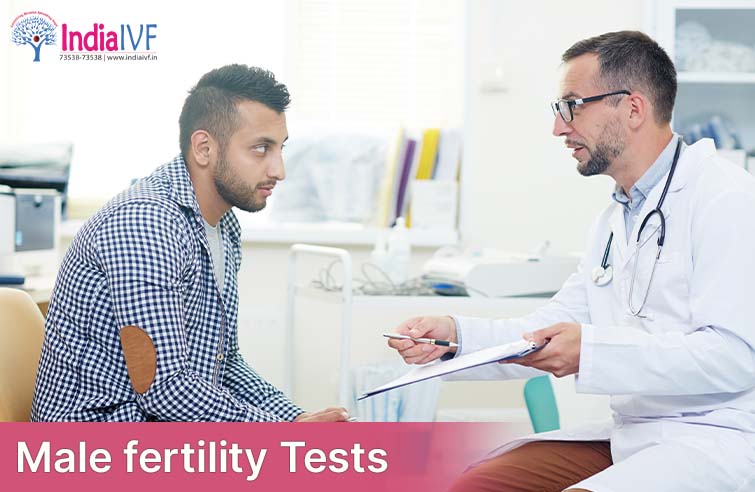
Male fertility tests play a crucial role in identifying issues affecting a man’s ability to father a child. They help couples struggling to conceive understand the underlying problems and create a personalized treatment plan. These tests offer valuable insights and can provide hope for many couples on their journey to parenthood.
If a couple is under 35 and has been trying to conceive through regular, unprotected intercourse for over a year without success, it’s time to consider fertility testing. This time frame allows for natural fluctuations in fertility, but after a year, it’s essential to explore potential causes.

Men with known risk factors for infertility should consider testing. These factors include a history of infections (such as mumps, gonorrhea, or chlamydia), varicocele, exposure to toxins or radiation, and hormonal imbalances. Being proactive can help address issues early on.
If a man has experienced previous fertility problems, such as unsuccessful pregnancies or a history of low sperm count or quality, it’s a good idea to undergo testing. This information can help doctors create the most effective treatment plan.
Sometimes, men may have anxiety or doubts about their fertility. In these cases, tests can provide peace of mind and confirm reproductive health.
Another essential aspect of male infertility evaluation is the general physical examination and medical history. These steps help healthcare professionals identify potential causes of infertility and better understand a patient’s overall health.
During the general physical examination, the healthcare provider will:
A comprehensive medical history helps identify potential factors contributing to infertility. The healthcare provider will ask about:
Lifestyle factors, such as exercise, diet, smoking, and alcohol consumption
By gathering information from the general physical examination and medical history, healthcare professionals can make informed decisions about the most appropriate diagnostic tests and treatment options for each individual patient.
Semen Analysis: Quantitative and Qualitative Assessment
Semen analysis is a comprehensive test that evaluates both quantitative (numerical) and qualitative (non-numerical) aspects of sperm. It is the primary diagnostic tool used to assess male fertility, providing valuable information about sperm count, motility, and morphology.
The World Health Organization (WHO) has established reference criteria for semen analysis parameters in their 2010 manual, “WHO Laboratory Manual for the Examination and Processing of Human Semen.” These criteria are widely accepted and provide a benchmark for evaluating male fertility.
The quantitative assessment involves measuring the following sperm parameters based on the latest WHO criteria:

Sperm motility: The percentage of sperm that move actively. The lower reference limit for total motility (progressive and non-progressive) is 40%.
Sperm concentration: The total number of sperm in the entire ejaculate. The lower reference limit is 39 million sperm/ejaculate.
The qualitative assessment evaluates sperm morphology (shape) and other aspects of the semen, based on the latest WHO criteria:
Sperm morphology: The percentage of sperm with a normal shape. The lower reference limit for normal forms is 4%.
Semen volume: The amount of semen produced during ejaculation. The lower reference limit is 1.5 mL.
Semen pH: A measure of the acidity or alkalinity of the semen. The normal pH range is 7.2 to 8.0.
Semen viscosity: The thickness or consistency of the semen. Normal semen should liquefy within 20-30 minutes after ejaculation.
| Parameter | Lower Reference Limit |
| Sperm count | 15 million/mLt |
| Total sperm motility | 40% |
| Sperm concentration | 39 million/ejaculate |
| Normal sperm morphology | 4% |
| Semen volume | 1.5 mL |
| Semen pH | 7.2-8.0 |
| Semen liquefaction time | 20-30 minutes |
Using the latest WHO reference criteria, healthcare professionals can accurately assess a man’s fertility potential and develop an appropriate treatment plan based on the semen analysis results.
A scrotal ultrasound is a non-invasive imaging test that uses high-frequency sound waves to create images of the scrotum and its contents. This painless procedure can help healthcare professionals evaluate the testicles, epididymis, and other structures within the scrotum, identifying any abnormalities that may contribute to male infertility.
Purpose of Scrotal Ultrasound
A scrotal ultrasound may be recommended for several reasons, including:
2. Procedure
A scrotal ultrasound typically involves the following steps:

3. Results
A radiologist or a healthcare professional will interpret the images from the scrotal ultrasound and provide a report outlining any findings. Depending on the results, further diagnostic tests or treatment options may be recommended.
Overall, a scrotal ultrasound is a valuable diagnostic tool in male fertility assessment, offering a safe and effective way to identify potential issues within the scrotum that may impact reproductive capabilities.
A transrectal ultrasound (TRUS) is a non-invasive imaging test that uses high-frequency sound waves to create images of the prostate gland and surrounding tissues. This diagnostic tool is particularly helpful in evaluating the male reproductive system, as it can detect abnormalities or obstructions that may contribute to infertility.

1. Purpose of Transrectal Ultrasound
A transrectal ultrasound may be recommended for several reasons, including:
2. Procedure
A transrectal ultrasound typically involves the following steps:
3. Results
A radiologist or healthcare professional will interpret the images from the transrectal ultrasound and provide a report outlining any findings. Depending on the results, further diagnostic tests or treatment options may be recommended.
In summary, a transrectal ultrasound is an essential diagnostic tool in male fertility assessment, offering a safe and effective method to identify potential issues within the prostate gland, ejaculatory ducts, and surrounding structures that may impact reproductive capabilities.
Hormone testing is an essential diagnostic tool in male fertility assessment, as hormonal imbalances can contribute to various reproductive issues, including low sperm count, poor sperm motility, and erectile dysfunction. Hormone testing helps healthcare professionals identify the underlying cause of these issues, guiding the development of appropriate treatment plans.
1. Hormones Involved in Male Fertility
Several hormones play essential roles in male fertility, including:
2. Hormone Testing Methods
Hormone testing typically involves a blood test, which measures the levels of hormones in the bloodstream. The test may be performed at various times, depending on the hormone being evaluated, such as:

3. Results
Interpretation of hormone test results depends on the hormone being evaluated and the patient’s age. For example, normal testosterone levels in men range from 300 to 1,000 nanograms per deciliter (ng/dL), while normal FSH levels range from 1.5 to 12.4 milli-international units per liter (mIU/mL) in men younger than 50 years old. In contrast, normal prolactin levels range from 2 to 18 ng/mL, regardless of age.
Abnormal hormone levels may indicate an underlying hormonal imbalance that can contribute to male infertility. Depending on the results, healthcare professionals may recommend further diagnostic tests, such as semen analysis or imaging studies, or initiate hormone therapy to address the underlying issue.
Post-ejaculation urinalysis is a diagnostic test that analyzes a urine sample collected immediately after ejaculation. This test is used to detect the presence of sperm in the urine, which can help healthcare professionals identify a condition called retrograde ejaculation, where sperm enters the bladder instead of being expelled through the urethra during ejaculation.

1. Purpose of Post-Ejaculation Urinalysis
A post-ejaculation urinalysis may be recommended for several reasons, including:
2. Procedure
A post-ejaculation urinalysis typically involves the following steps:
3. Results
If sperm are detected in the post-ejaculation urine sample, it may indicate retrograde ejaculation. Healthcare professionals will consider the test results along with the patient’s medical history, physical examination, and other diagnostic tests to determine the underlying cause and develop an appropriate treatment plan.
In some cases, retrograde ejaculation can be managed with medications that help close the bladder neck during ejaculation or by addressing the underlying cause, such as diabetes or nerve damage. In cases where fertility is affected, assisted reproductive techniques, such as in vitro fertilization (IVF) or intracytoplasmic sperm injection (ICSI), may be recommended.
In summary, post-ejaculation urinalysis is a useful diagnostic tool for detecting retrograde ejaculation and guiding treatment options. By analyzing the presence of sperm in the urine, healthcare professionals can gain insights into potential issues affecting fertility and develop appropriate treatment plans.
A testicular biopsy is a diagnostic procedure that involves the removal of a small tissue sample from one or both testicles. This test is typically performed when semen analysis reveals no sperm (azoospermia) or extremely low sperm counts (severe oligozoospermia) to determine the cause of the issue and identify the most appropriate treatment options.
1. Purpose of Testicular Biopsy
A testicular biopsy may be recommended for several reasons, including:
2. Procedure
A testicular biopsy can be performed using one of the following techniques:

Both methods are typically performed under local anesthesia, with sedation or general anesthesia being an option in some cases. The procedure usually takes around 30-60 minutes, and the patient may experience mild discomfort, swelling, or bruising afterward.
3. Results
The extracted testicular tissue is analyzed in a laboratory by a pathologist, who will evaluate the sample for sperm production, the presence of sperm, and any abnormalities in the tissue. Depending on the results, healthcare professionals may recommend further diagnostic tests, treatments, or assisted reproductive techniques to address the underlying cause of infertility.
In summary, a testicular biopsy is a valuable diagnostic tool for identifying the causes of male infertility, particularly when semen analysis results are inconclusive. By examining testicular tissue, healthcare professionals can gain insights into sperm production and potential issues affecting fertility, guiding the development of appropriate treatment plans.
While semen analysis provides valuable information about sperm count, motility, and morphology, it does not always identify specific issues affecting sperm function. Specialized sperm function tests are designed to evaluate various aspects of sperm performance, which can be crucial for successful fertilization and pregnancy. These tests can help healthcare professionals understand the underlying causes of male infertility and guide treatment options.

While semen analysis provides valuable information about sperm count, motility, and morphology, it does not always identify specific issues affecting sperm function. Specialized sperm function tests are designed to evaluate various aspects of sperm performance, which can be crucial for successful fertilization and pregnancy. These tests can help healthcare professionals understand the underlying causes of male infertility and guide treatment options.
The sperm penetration assay (SPA), also known as the hamster egg penetration test, assesses the ability of sperm to penetrate and fertilize an egg. In this test, sperm are incubated with hamster eggs that have been stripped of their zona pellucida (outer shell). The test measures the number of eggs penetrated by sperm, providing an indication of fertilization potential.
The hypoosmotic swelling test evaluates the integrity of the sperm membrane, which is essential for successful fertilization. Sperm are exposed to a hypoosmotic solution, causing the sperm with intact membranes to swell and curl. The percentage of sperm that reacts to the solution is indicative of membrane functionality.
This test measures the levels of reactive oxygen species (ROS) in semen, which are molecules produced as a byproduct of normal cellular metabolism. High levels of ROS can cause oxidative stress, potentially damaging sperm DNA, membranes, and other cellular components. The ROS test helps identify cases where oxidative stress may be contributing to infertility.
The sperm DNA fragmentation test assesses the integrity of sperm DNA, which is vital for the development of a healthy embryo. This test uses a variety of techniques, such as the sperm chromatin dispersion test (SCD) or the terminal deoxynucleotidyl transferase dUTP nick end labeling (TUNEL) assay, to detect breaks or damage in the sperm DNA.
In some cases, a man’s immune system may produce antibodies that target and impair sperm function, leading to infertility. The antisperm antibody test detects the presence of these antibodies in the semen, helping healthcare professionals identify cases where an immune response may be affecting fertility. While specialized sperm function tests are not typically performed as part of routine fertility assessments, they can be valuable diagnostic tools when conventional semen analysis fails to explain fertility issues. These tests provide further insights into sperm function and potential causes of infertility, guiding healthcare professionals in creating effective treatment plans.
A male fertility test may be recommended if a couple has been actively trying to conceive for 12 months or more without success, or if there are any known risk factors for male infertility, such as previous testicular surgery, chemotherapy or radiation therapy, or certain medical conditions.
While a blood test cannot directly detect male infertility, it can identify hormonal imbalances or medical conditions that may contribute to infertility, such as low testosterone levels or thyroid dysfunction.
A sperm fertility test, or semen analysis, typically takes around 1-2 hours to complete, including sample collection and analysis.
The ideal time for a sperm test is 2-7 days after the last ejaculation, as this allows for sufficient semen volume and optimal sperm motility.
At India IVF Clinics we provide the most comprehensive range of services to cover all the requirements at a Fertility clinic including in-house lab, consultations & treatments.
