In the world of modern medicine, women’s health has taken a front seat. One of the most essential tools used by doctors to monitor and diagnose issues within the female body is the pelvic ultrasound. This safe and non-invasive procedure allows doctors to look inside the body without making any cuts. Whether a woman is experiencing unusual pain, irregular periods, or is simply checking on her pregnancy, a pelvic ultrasound provides the clarity needed to make the right medical decisions. Understanding how this technology works and why it is used can help ease any anxiety you might have about the procedure.
What is a Pelvic Ultrasound?
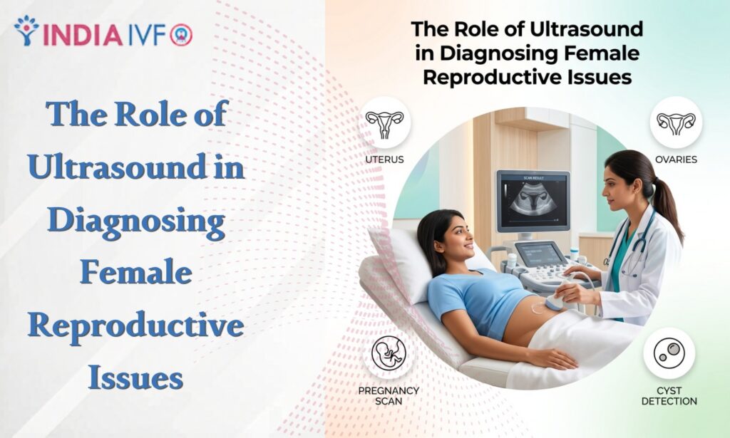
A pelvic ultrasound is a diagnostic imaging test that uses high-frequency sound waves to create a picture of the organs inside your pelvis. These organs include the uterus, cervix, vagina, fallopian tubes, and ovaries. Unlike X-rays, an ultrasound does not use radiation, making it very safe for women of all ages, including those who are pregnant.
When you go for a pelvic ultrasound, a small device called a transducer is used. This device sends out sound waves that bounce off your internal organs. These echoes are then sent back to a computer, which turns them into a real-time image on a screen. It is a fundamental part of reproductive health imaging because it allows doctors to see the size, shape, and condition of your reproductive organs instantly.
The Different Types of Pelvic Ultrasound
When your doctor orders a pelvic ultrasound, they may choose one of two main methods, or sometimes both, depending on what they need to see.
1. Transabdominal Ultrasound
In this method, the transducer is moved across the skin of your lower abdomen. Usually, you are asked to have a full bladder for this test. The water in your bladder acts like a window, helping the sound waves travel more clearly so the doctor can get a better view of the uterus and ovaries.
2. Transvaginal Scan
A transvaginal scan is often more detailed than an abdominal one. In this procedure, a specially designed thin transducer is covered with a sterile sheath and inserted into the vagina. While it might sound intimidating, it is usually not painful and only involves a bit of pressure. This method is excellent for getting a close-up look at the lining of the uterus and the ovaries, which is why it is frequently used for ovarian cysts diagnosis and checking early pregnancies.
Why Do Doctors Recommend a Pelvic Ultrasound?
There are many reasons why a woman might need a pelvic ultrasound. It is the first line of defense when something feels “off” in the reproductive system.
- Identifying the Cause of Pelvic Pain: If you are experiencing unexplained pain in your lower stomach, a pelvic ultrasound can help find the source, such as inflammation or infection.
- Investigating Abnormal Bleeding: If your periods are very heavy or if you are bleeding between periods, the doctor uses imaging to see if there are growths inside the uterus.
- Monitoring Pregnancy: This is the most well-known use. It helps check the baby’s heartbeat, growth, and position.
- Checking the Position of an IUD: If you use an intrauterine device for birth control, a pelvic ultrasound can ensure it is placed correctly.
- Fertility Evaluations: For women struggling to conceive, imaging helps track the growth of eggs in the ovaries.
Ovarian Cysts Diagnosis through Pelvic Ultrasound
One of the most common findings during a pelvic ultrasound is the presence of ovarian cysts. Ovarian cysts are fluid-filled sacs that develop on or inside the ovaries. Most women will develop at least one cyst during their lifetime, often without even knowing it.
Using a pelvic ultrasound, a doctor can perform an accurate ovarian cysts diagnosis. The imaging shows whether the cyst is filled with fluid (a simple cyst) or if it contains solid material (a complex cyst). Simple cysts are usually harmless and often go away on their own. However, if the ultrasound shows a complex cyst, the doctor may need to perform further tests to rule out more serious conditions.
Detecting Fibroids and Other Uterine Issues
Fibroids are non-cancerous growths that develop in or on the muscular wall of the uterus. They are very common and can cause symptoms like heavy bleeding, pelvic pressure, and frequent urination.
A pelvic ultrasound is the best way to detect fibroids. The scan shows the doctor exactly where the fibroids are located and how large they have grown. This information is crucial for deciding on a treatment plan, whether it involves medication or a minor surgical procedure. Without reproductive health imaging, it would be very difficult to know why a woman is experiencing such heavy and painful periods.
The Importance of the Transvaginal Scan in Early Diagnosis
In many cases, a standard scan over the stomach isn’t enough to see the fine details. This is where the transvaginal scan becomes a hero. Because the probe is closer to the internal organs, the images are much sharper.
This type of scan is particularly useful for:
- Diagnosing ectopic pregnancies (when the embryo grows outside the uterus).
- Checking the thickness of the endometrial lining.
- Finding small polyps that might be causing bleeding.
- Early detection of cancers in the reproductive system.
By using a transvaginal scan, doctors can catch issues much earlier than they would with a physical exam alone.
How Reproductive Health Imaging Supports Fertility
For many women, the journey to motherhood is not always easy. Reproductive health imaging plays a vital role in fertility treatments. A pelvic ultrasound is used to perform “follicle tracking.” This means the doctor monitors the ovaries to see when eggs are maturing and when ovulation is about to occur.
Additionally, imaging can check for “hydrosalpinx,” which is a condition where the fallopian tubes are blocked with fluid. Identifying these issues early through a pelvic ultrasound can save a woman months or even years of frustration when trying to get pregnant.
What to Expect During Your Pelvic Ultrasound Appointment
If you have never had a pelvic ultrasound before, you might feel a bit nervous. Here is a simple breakdown of what usually happens:
- Preparation: For an abdominal scan, you will be told to drink 32 ounces of water an hour before the test. For a transvaginal scan, you will be asked to empty your bladder right before the test starts.
- The Procedure: You will lie on an exam table. A clear gel will be applied to your skin (for abdominal) or the probe (for transvaginal). This gel helps the sound waves travel.
- The Scan: The Doctor will move the transducer to get the best images. You might feel some pressure, but it should not be painful.
- After the Test: You can wipe off the gel and go about your day immediately. There is no “recovery time” needed for a pelvic ultrasound.
The Benefits of Pelvic Ultrasound Over Other Methods
While there are other tests like CT scans or MRIs, the pelvic ultrasound remains the preferred choice for many reasons:
- Safety: No radiation is used. This is why it is safe for pregnant women and developing babies.
- Cost-Effective: It is generally much cheaper than an MRI or a CT scan.
- Speed: The results are often available quickly, and the procedure itself takes only 20 to 30 minutes.
- Real-time Results: Doctors can see the organs moving in real-time, such as a baby moving or blood flowing through vessels.
Risks and Limitations of Pelvic Ultrasound
While a pelvic ultrasound is incredibly safe, it is important to understand its risks and limitations.
Risks:
-
No Known Biological Risks: Since it uses sound waves instead of radiation, there are no known long-term risks to the patient or a fetus.
-
Discomfort: The main “risk” is minor physical discomfort. A full bladder (required for abdominal scans) can be uncomfortable, and the insertion of a probe for a transvaginal scan can cause a sensation of pressure.
Limitations:
-
Body Habitus: In some cases, intestinal gas or excess abdominal fat can make it difficult for sound waves to pass through, resulting in less clear images.
-
Need for Further Testing: While a pelvic ultrasound is excellent for finding growths, it cannot always determine if a growth is cancerous or benign. In such cases, an MRI or a biopsy may be needed.
Cost and Comparison Table
Below is a general comparison of common pelvic imaging procedures.
| Feature | Transabdominal Ultrasound | Transvaginal Scan | Pelvic MRI |
| Primary Use | General overview, Pregnancy | Detailed view of Ovaries/Uterus | Complex cases, Cancer staging |
| Preparation | Full Bladder required | Empty Bladder required | Varies (sometimes fasting) |
| Radiation | None | None | None |
| Average Cost | ₹18,800 – ₹47,000 | ₹23,500 – ₹56,400 | ₹94,000 – ₹2,82,000 |
| Accuracy | High (for large masses) | Excellent (for small details) | Very High |
Understanding Common Conditions Found via Pelvic Ultrasound
Beyond fibroids and ovarian cysts diagnosis, a pelvic ultrasound can help identify several other conditions:
Endometriosis
While a pelvic ultrasound cannot always see small spots of endometriosis, it is very good at finding “endometriomas,” which are cysts on the ovaries caused by this condition.
Polycystic Ovary Syndrome (PCOS)
PCOS is a common hormonal disorder. On a pelvic ultrasound, a doctor can see if the ovaries have a “string of pearls” appearance, which is a sign of many small follicles that haven’t developed into eggs.
Pelvic Inflammatory Disease (PID)
This is an infection of the reproductive organs. Imaging can show if there is fluid in the fallopian tubes or abscesses in the pelvis, helping the doctor start the right antibiotics quickly.
The Future of Reproductive Health Imaging
Technology is always improving. Today, we have 3D and 4D pelvic ultrasound options. These provide even more detailed views, allowing doctors to see the shape of the uterus in three dimensions. This is particularly helpful for women who have had multiple miscarriages, as it can reveal structural issues in the uterus that a 2D scan might miss.
As reproductive health imaging continues to advance, the ability to diagnose issues early and accurately only gets better. This means less invasive surgeries and more personalized care for women.
Conclusion: Taking Charge of Your Health
The pelvic ultrasound is an incredible tool that has changed the way women’s healthcare is delivered. It takes the guesswork out of diagnosing pain, bleeding, and fertility issues. By providing a clear window into the body, a pelvic ultrasound ensures that conditions like fibroids and ovarian cysts are found and treated early. If your doctor recommends a transvaginal scan or a standard abdominal scan, remember that it is a step toward understanding your body better. It is a painless, safe, and highly effective way to prioritize your reproductive health. Regular check-ups and the right imaging can make all the difference in living a healthy, pain-free life.